Imaging of Unusual Ingested Multiple Stones in a Child: A Case Report
Osward Bwanga
Midlands University Hospital Tullamore, Radiology Department, Co. Offaly, Ireland
Mulewa Mulenga
Arthur Davison Children’s Hospital and Copperbelt University, Ndola
Ethel Chilambe Jere
Arthur Davison Children’s Hospital, Radiology Department, Ndola
Luyando Simunyama
Arthur Davison Children’s Hospital and Copperbelt University, Ndola
Patrick Jila
Arthur Davison Children’s Hospital, Surgery Department, Ndola
Abraham Siingwa
Zambia Flying Doctor Services, Radiology Department, Ndola
DOI: https://doi.org/10.55320/mjz.52.1.621
Keywords:Geophagia, Imaging, Ingested foreign body, Plain film radiography, Zambia
ABSTRACT
Ingested foreign bodies (FBs) in children are a common referral for imaging examinations in paediatric hospitals. Most ingested FBs pass without any intervention or complications. In a few cases, they can cause significant complications such as obstruction, bleeding, perforation, fistulisation, sepsis and death. Therefore, early patient presentation to the medical facility, diagnosis and management are crucial to avoid associated complications and death. Imaging plays an essential role in diagnosing, monitoring and treating ingested FBs. Most ingested radiopaque FBs are identified using plain film radiography. In the case of radiolucent FBs and complications, ultrasound and computed tomography (CT) imaging examinations are used. In this case report, we report an unusual imaging of a 2-year-old boy who was diagnosed with multiple stones in the colon and rectum. Without clinical information on the radiology request form (RRF) and the absence of the radiologist, the radiographic appearance of multiple stones in the colon and rectum could easily be mistaken for residual barium sulphate from barium studies. This dilemma shows the importance of correlating radiographic features with clinical findings during radiographic image interpretation.
INTRODUCTION
Foreign body (FB) ingestion in children is common and mainly observed in children between 6 months and 3 years old.[1-3] Children's curiosity and need to explore the world around them place them at a higher risk of ingesting FBs.[4] The most common FBs ingested by children include coins, toy parts, dentures, magnets, batteries, and needles.[1][4][5] These FBs may be swallowed accidentally or occasionally intentionally.[5] Children can also intentionally ingest soil, gravel, clay, or stones, a condition called geophagia.[6][7] The deliberate ingestion of FBs is rare in older children and healthy adults but is frequently seen in psychiatric patients and patients with additional intellectual needs.
The ingestion of FBs is a significant problem in children.[1][3] Approximately 80% to 90% of FBs in the gastrointestinal tract (GIT) are passed spontaneously via faecal matter without complications, 10% to 20% are removed endoscopically, and 1% require open surgery secondary to complications.[3] If the FB is lodged in the oesophagus or other parts of the GIT, significant complications such as aspiration, obstruction, bleeding, perforation, fistulisation, sepsis, and death may occur.[3][4][8] In the United States of America (USA), 1500 people die each year due to ingested FBs.[4][9] Therefore, early patient presentation to the medical facility, initial assessment, diagnosis, and management are crucial to avoid associated complications and death.
The initial patient assessment aims to identify the type of FB ingested, its location in the GIT, and any associated complications.[3][4] This role is mainly performed using medical imaging examinations. In radiology departments, the common imaged (X-rayed) ingested radiopaque FBs are coins, toy parts, magnets, batteries, dentures, and needles. However, imaging of ingested stones is unusual. In this imaging case report, we report an unusual case of a 2-year-old boy who presented to our teaching hospital with painful bowel movements, difficulty passing stool, and the passage of multiple stones. To the best of the authors' knowledge, this is the first case report on geophagia to be documented from the medical imaging perspective in Zambia and globally. A few documented case reports on geophagia are from the medical and surgical perspectives.[10-12] Therefore, this case report focuses on medical imaging.
CASE PRESENTATION
This is an imaging case report of a 2-year-old boy who presented to our teaching hospital with difficulty passing stool, painful bowel movements, and the passage of hard stones for a day. The child had been well until the past month when her mother had gone to work in another town and left him in the care of a relative. Upon the mother's return, she noticed that the child was having difficulties opening his bowels and would cry whenever he tried to do so. The mother observed blood-stained stones coming out of the child's anus as he strained to defecate. This prompted the mother to take the child to a nearby clinic, where he was referred to our teaching hospital. The child's mother reported that he started picking up and eating sand from the ground when he was one year old. The mother also suspected that the stones the child was passing might be construction stones that a neighbour had obtained for home renovation. There was no history of psychiatric or intellectual needs for the child. The medical practitioners requested an abdominal X-ray examination to confirm the ingestion of the stones and rule out associated complications.
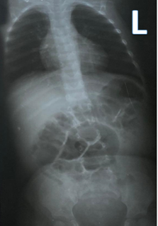
The paediatric patient arrived at the radiology department wearing a hospital gown and was accompanied by the mother and nurse. The procedure was explained, and the patient and X-ray equipment were prepared. The mother was asked to wear the lead apron and hold the child during the examination. A single supine anteroposterior (AP) projection of the chest and abdomen was undertaken using a 35 by 43 centimetres film-screen cassette. The X-ray film was processed, and Figure 1 shows the resultant initial radiograph.

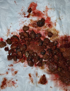
The radiograph showed multiple rounded radiopaque densities throughout the colon and rectum, consistent with multiple stones. The differential diagnosis was residual barium sulphate from barium studies. However, there was no recent history of barium study. The child was admitted to the hospital and treated with lactulose to soften the stool. The stones were removed using non-operative management, which involved instrumental removal, diet, and enemas to facilitate natural defecation. After two days of treatment, a repeat abdominal X-ray was requested and performed, which showed no stones (Figure 2). Figure 3 also shows the stones removed and defecated by the paediatric patient.
The referring medical practitioner interpreted the radiographs because our teaching hospital does not have a radiologist. There were no associated complications, and the patient was discharged on the fifth day of admission. No further follow-up imaging was required.
DISCUSSION
The imaging process starts with the referring medical practitioner requesting the examination by completing the radiology request form (RRF). In the case of suspected ingested FBs, the approximate time of swallowing the object(s) and the site of any localised discomfort should be ascertained and noted on the initial RRF, along with the time the imaging examination is undertaken.[5] This information allows the radiographer to determine the body part to be imaged and the best radiographic projections to be conducted. The FB locations may change after the imaging examination. Therefore, indicating the time of the imaging examination by the radiographer is helpful to the referring medical practitioner. Unfortunately, this information was missing from our case report on the initial RRF.
In this case report, plain film radiography was used to diagnose and monitor ingested multiple stones. This corresponds with the literature, which revealed that plain film radiography is the most used imaging examination to diagnose ingested radiopaque FBs.[4][5] This medical imaging method can confirm the location, size, shape, and number of ingested radiopaque FBs and help exclude aspiration.[9] It is also instrumental in excluding complications associated with ingested FBs, such as the presence of pneumoperitoneum, bowel obstruction, and faecal impaction.[8] Other advantages of plain film radiography include availability, affordability, simplicity, and low radiation dose.[5][13][14] If the patient is a young child, the imaging examination is usually restricted to a single anteroposterior projection, including the neck, chest, and abdomen.[5] This conforms with what was undertaken in this case report (Figures 1 and 2).
Plain film radiography may be insufficient in identifying ingested radiolucent FB and associated complications.[9][13][15] Ingested radiolucent FBs may be detected using oral contrast media administration, such as barium sulphate. However, oral contrast media may sometimes obscure the FBs.[9] The use of barium sulphate should be avoided if there is any suspicion of perforation because of the potential for contrast spillage into the mediastinum or pleural space.[9][16] In such cases, water-soluble, iodinated contrast medium such as gastrografin should be used instead of barium sulphate.[16][17] Ultrasound imaging is also an alternative imaging examination for detecting ingested radiolucent FBs and may be used to locate and track the FBs as they pass through the GIT.[15] It is inexpensive and has the advantage of not using ionising radiation.[15][18] Computed tomography (CT) is another imaging examination used to detect and determine the location and structure of ingested FBs. Additionally, CT is useful in assessing complications of FBs because of its capabilities, such as showing detailed anatomical and enabling multiplanar examination.[13] Therefore, imaging examinations complement each other in detecting ingested FBs and associated complications.
In our case report, the RRF had clinical information that helped in imaging the patient and interpreting the radiographic images. Without such clinical information and with the radiologist's current absence, the appearance of multiple stones in the colon and rectum (Figure 1) might easily have been mistaken for residual barium sulphate from barium studies.
Our teaching hospital has no radiologist to interpret and report on radiographic images. The Ministry of Health (MOH)[19] has identified a critical shortage of radiologists as one of the challenges facing the delivery of quality imaging services in Zambia. At the time of writing this case report, 22 radiologists were serving a population of 19.6 million.[20] This has resulted in the attraction of a few radiologists towards advanced imaging examinations such as CT and magnetic resource imaging (MRI), leaving the interpretation of plain radiographs to the referring medical practitioners,[21] despite their limited training in this area.[22-24] This may lead to misdiagnosis and mismanagement of patients.[23-27] To overcome this challenge, some countries, such as the UK, Denmark, and Uganda, have established reporting radiographers to fill the gap created by the shortage of radiologists. To improve our radiology reporting services, the Radiological Society of Zambia (RSZ) needs to engage stakeholders and advocate for reporting radiographers. The Health Profession Council of Zambia (HPCZ) should extend the scope of professional practice for radiographers and schools of radiography to establish image interpretation and reporting postgraduate courses.
CONCLUSION
Ingested FBs in children are a common referral for imaging examinations. Radiographers commonly X-ray children with ingested FBs such as coins, toy parts, magnets, batteries, and needles, but stones are rare. In this case report, plain film radiography was the main imaging examination used in the diagnosis and management of ingested multiple stones in the colon and rectum due to its advantages of being easy to access, low cost, and low radiation dose. The stones were removed using non-operative management, which involved instrumental removal, diet, and enemas to facilitate natural defecation. This case report shows the role and importance of medical imaging in diagnosing and managing ingested FBs.
CONSENT FOR PUBLICATIONThe authors obtained written and signed informed consent from the child's mother for the publication of this case report and accompanying radiographic images and photo of stones. Permission was also obtained from the Senior Medical Superintendent to use radiographic images and patient information for the case report publication.
REFERENCES
- Dereci S, Koca T, Serdaroğlu F, Akçam M. Foreign body ingestion in children. Turk Pediatri Ars. 2015;50(4):234-240. Published 2015 Dec 1. doi:10.5152/TurkPediatriArs.2015.3164
- Lee JH. Foreign Body Ingestion in children. Clin Endosc. 2018;51(2):129-136. doi:10.5946/ce.2018.039
- Gatto A, Capossela L, Ferretti S, et al. Foreign body ingestion in children: epidemiological, clinical features and outcome in a third level emergency department. Children (Basel). 2021;8(12):1182. Published 2021 Dec 15. doi:10.3390/children8121182
- Pinto A, Lanza C, Pinto F, et al. Role of plain radiography in the assessment of ingested foreign bodies in the pediatric patients. Semin Ultrasound CT MR. 2015;36(1):21-27. doi:10.1053/j.sult.2014.10.008
- Whitley AS, Jefferson G, Sloane KHC, Anderson G, Hoadley G. Clark’s positioning in radiography (13th ed). London: CRC Press Ltd; 2015.
- Bonglaisin JN, Kunsoan NB, Bonny P, et al. Geophagia: Benefits and potential toxicity to human-A review. Front Public Health. 2022;10:893831. Published 2022 Jul 26. doi:10.3389/fpubh.2022.893831
- Ranchod AI. Geophagy; 2023. From https://radiopaedia.org/articles/geophagy (Accessed 15 December 2024)
- Clarke CGD, Dux AEW. Abdominal X‐rays for medical students. West Sussex: John Wiley & Sons, Ltd; 2015.
- Guelfguat M, Kaplinskiy V, Reddy SH, DiPoce J. Clinical guidelines for imaging and reporting ingested foreign bodies. AJR Am J Roentgenol. 2014;203(1):37-53. doi:10.2214/AJR.13.12185
- Narayanan PV, Balachandran MK. Colonic obstruction after ingested gravel and stone. Asian J Surg. 2012;35(2):96-98. doi:10.1016/j.asjsur.2012.04.02
- Malik AM. Deliberate ingestion of stones causing a diagnostic dilemma. A personal experience. Int J Health Sci (Qassim). 2015;9(1):83-86.
- Hans AK, Brown JM, Qayed E. A massive stone ingestion. ACG Case Rep J. 2021;8(11):e00707. Published 2021 Nov 23. doi:10.14309/crj.0000000000000707
- Deniz MA, Turmak M. CT evaluation of swallowed foreign bodies located in the gastrointestinal system. Cureus. 2022;14(6):e26355. Published 2022 Jun 26. doi:10.7759/cureus.26355
- Halim NI, Mohd Zaki F, Manan HA, Mohamed Z. An evaluation of the quality of plain radiograph interpretations by radiology trainees: A single institution experience. Diagnostics (Basel). 2022;12(8):1954. Published 2022 Aug 12. doi:10.3390/diagnostics12081954
- Wong BM, Wong SR, Nesiri C, Udayasankar U, Larson MC. Ultrasound imaging of various ingested foreign bodies in an Ex Vivo Intestinal Model. Pediatr Emerg Care. 2024;40(12):850-855. doi:10.1097/PEC.0000000000003269
- Bontrager KL, Lampignano JP. Radiographic positioning and related anatomy (6th ed). Mosby: MI. St. Louis; 2014.
- Bwanga O, Chanda E, Mwansa E, Nkoloma B. Changing roles of a radiographer in fluoroscopy: literature review to guide the development and practice of fluoroscopic studies in Zambia. Medical Journal of Zambia. 2023; 49(4): 342-351.
- Bwanga O, Mwase G, Kaunda HC. Midwives’ experiences of performing obstetric ultrasounds in maternity care: a systematic review. African Journal of Midwifery and Women’s Health. 2021; 15(2): 1-8.
- Ministry of Health. National health strategic plan 2022-2026. Lusaka: The Ministry of Health; 2023.
- Mubanga B, Bwanga O, Sichone J, Kafwimbi S, Sichone PN. Education and training in intravenous cannulation and administration of contrast media in Radiography: A literature review. Medical Journal of Zambia. 2024; 51(4): 381-391. doi:10.55320/mjz.51.4.592
- Bwanga O, Mulenga J, Chanda E. Need for image reporting by radiographers in Zambia. Medical Journal of Zambia. 2019; 46(3): 215-220. doi:10.55320/mjz.46.3.223
- Karera A, Engel-Hills P, Davidson F. Radiology image interpretation services in a low-resource setting: Medical doctors' experiences and the potential role of radiographers. Radiography. 2024;30(2):560-566. doi:10.1016/j.radi.2024.01.009
- Bwanga O, Chinene B, Kapapa N. Referring medical practitioners’ experiences with medical imaging services: Literature review findings and implications for Zambia. Medical Journal of Zambia. 2024; 51(4): 392-404. doi:10.55320/mjz.51.4.607
- Gam NP, Sibiya MN. Doctors' perspectives on the quality of medical imaging in public hospitals in eThekwini District. Health SA. 2024;29:2389. Published 2024 May 7. doi:10.4102/hsag.v29i0.2389
- Bwanga O, Chanda E, Kafwimbi S, Sichone J. Opinions of Zambian radiographers on extending their role in the interpretation and reporting on general radiographic images: a cross-sectional survey. Medical Journal of Zambia. 2021; 48(3): 212-220.
- Chilambe E, Muller H, du Plessis J. Novel training approach to improve a cohort of radiographers' image interpretation skills of trauma chest radiographs. J Med Imaging Radiat Sci. 2024;55(2):244-257. doi:10.1016/j.jmir.2024.02.003
- Bwanga O, Sichone J, Sichone P, Kazuma Y. Image interpretation and reporting by radiographers in Africa: Findings from the literature review and their application to Zambia. Medical Journal of Zambia. 2021; 48(2):125-135.
Medical Journal of Zambia, Vol 52, 1
The Medical Journal of Zambia, ISSN 0047-651X, is published by the Zambia Medical Association.
© This is an Open Access article distributed under the terms of the Creative Commons Attribution License, which permits unrestricted use, distribution, and reproduction in any medium, provided the original work is properly cited.